0.4 Tesla standing equine MRI
Stand.
Scan.
Know.
The standing MRI, reimagined at 0.4T, with a console your team runs from an iPad at the horse's side.
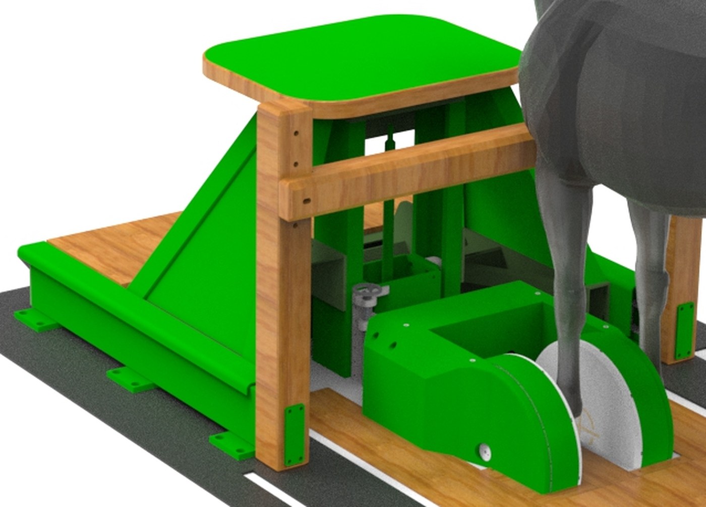
No general anaesthesia. A sedated horse walks onto the platform, the magnet is positioned at the limb, and the protocol AutoRuns. Motion is continuously monitored and corrected. Images reconstruct on a GPU with deep-learning denoising and appear in the viewer before the horse leaves the room.
- 0.4 T
- Field strength
- Continuous
- Motion tracking
- ≤ 5 s
- Between scans
- 0
- Anaesthetics
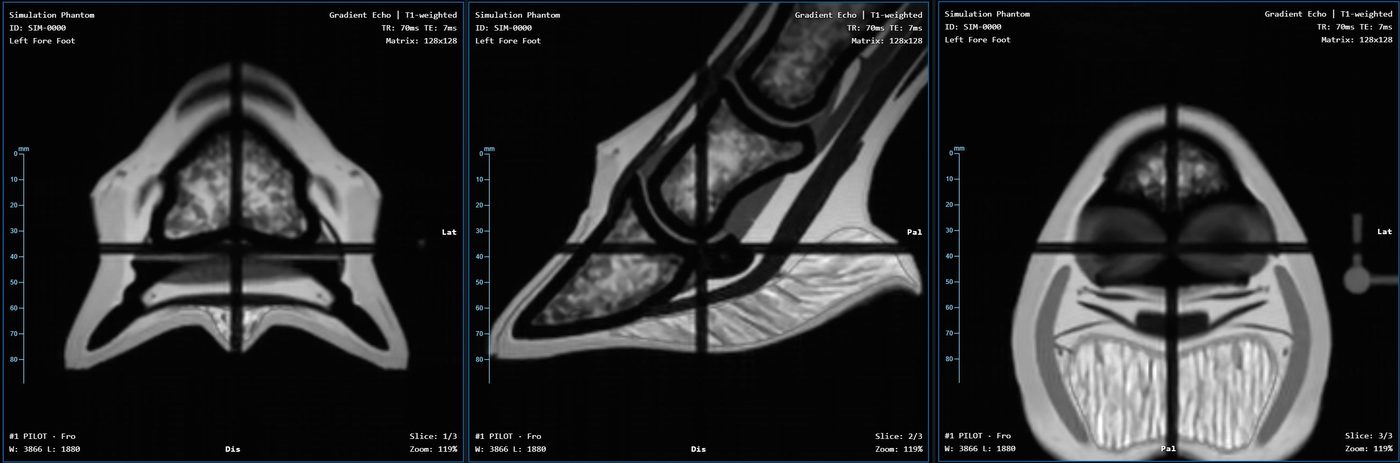
From the scanner, 9 November 2026
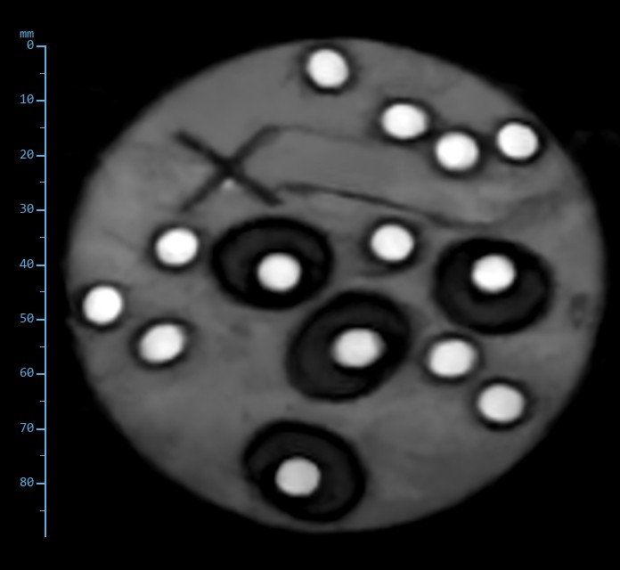
One phantom, six contrasts
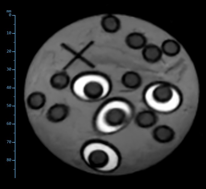
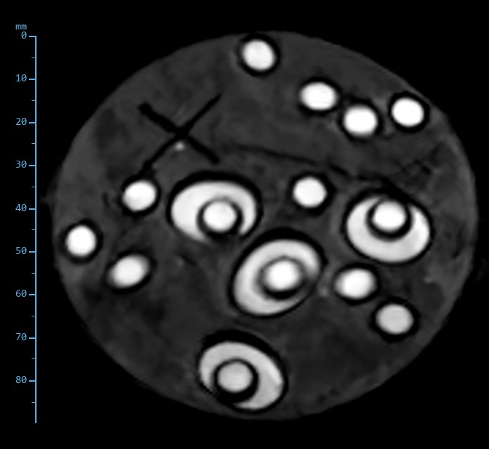
Pilot, proton density, T1, T2, T2* and STIR on a three-media phantom, straight from the console. Fluid, fat and muscle analogues rank the way they do in the limb. Then the same protocol on a pork hock, a grapefruit, a pomegranate and three duck eggs.
Why it is different
Four things changed
Standing MRI removed the anaesthetic from equine imaging two decades ago. The magnets, electronics and software behind the systems in service today have barely moved since. AltraMRI starts again with what is possible now.
Steadier
A sedated horse sways. AltraMRI is built around comprehensive motion compensation: the limb is tracked continuously through the whole of every scan, and the data taken during the acquisition corrects it.
- Tracked continuously, not sampled a few times a second
- Independent of the pulse sequence, and applies to every family
- Makes 3D practical on a standing horse: finer resolution and more signal, no longer spoiled by sway
Sharper
A 0.4T permanent magnet, roughly half again the field of a 0.27T standing system, with an integral transmit coil for a uniform B1 field. More signal in every voxel, spent on thinner slices, shorter scans, or both.
- Spin echo, fast spin echo, STIR and gradient echo families, 2D and 3D
- Even signal across the whole field of view from the integral transmit coil
- Deep-learning denoising and Gibbs-ringing suppression on the complex raw data, at native resolution
- Higher-resolution and fast variants of every family
Faster
Every scan takes its own pre-shot and centres itself, before and after each scan. Queue the exam and start it; the system runs the protocol with five seconds or less between scans.
- 0.4 T gives faster scanning for the same images
- Calibration runs on the first Start, with one-tap defaults for repeat patients
- Live progress and time remaining on every connected screen
Simpler
A web application, not a console PC in another room. Open it on an iPad or laptop anywhere on the clinic network. Nothing to install.
- Exam, Scan, Images and Archive a tap apart
- Operator, Clinical and Administrator roles with a locked clinical pipeline
- Synthetic exam mode for training and demonstrations without the magnet
Motion
Built for a horse that moves
Below the fetlock the hoof sits in the coil and movement is small. Above it, sway is the main reason a standing scan is repeated or a region is called non-diagnostic. Motion correction is not a feature on AltraMRI. It is part of the architecture.
Standing systems in service today
- How motion is measured
- Extra echoes inside the fast spin echo train
- How often
- Three position samples per second
- Which scans
- Fast spin echo and STIR; other scan types not corrected
From the manufacturer's published description, 2025.
AltraMRI
- How motion is measured
- Tracked independently of the echo train
- How often
- Continuously, for the whole scan
- Which scans
- Every family: spin echo, fast spin echo, STIR, gradient echo, 2D and 3D
The workflow protects the exam as well: every image reconstructs on the spot, and any scan can be repeated before the horse leaves the platform.
A day-patient exam
How a scan runs
- 01
Arrive
Shoes off, nail-fragment check, light sedation. A handler stays at the head throughout.
- 02
Position
The horse walks onto the platform. The Magnet Handling Unit positions the magnet to the limb.
- 03
Pilot
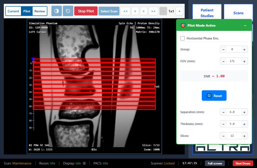
A three-plane locator in under a minute. Draw the slices you want, at any angle, and the scan is acquired where you piloted them.
- 04
Scan
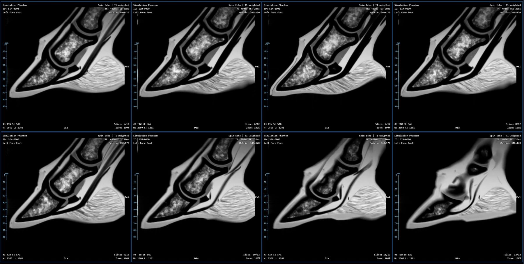
The protocol runs itself: T2 fast spin echo in three planes, STIR, proton density and T1 as the case requires. Repeat any scan on the spot.
- 05
Review
Images appear in the viewer as each scan reconstructs. Export as DICOM to your radiologist. The horse goes home the same day.
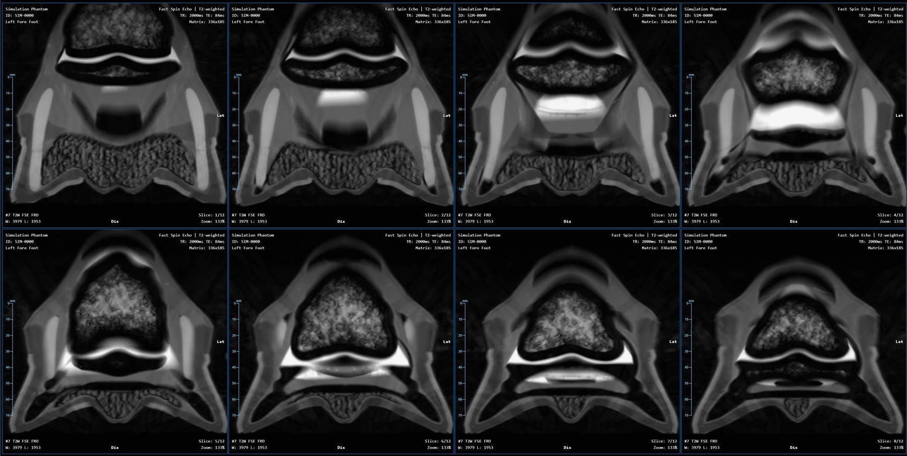
The console
Run the whole exam from the horse's side
- Scans that centre themselvesEvery technique sets its own frequency from a pre-shot, before and after each scan. Calibration runs on the first Start. Between scans the system is ready in five seconds or less.
- Draw it, scan itPlan slices on the pilot at any angle and position, including double-oblique. The scan lands exactly where the line was drawn.
- Protocols that runQueue a whole exam and start it. Abort one scan and the next begins. Progress and time remaining update live.
- Images on the spotA Cornerstone3D viewer with window, zoom, stack scrolling and three-plane review, in the same screen as the scan queue.
- Your images, your radiologistStandard DICOM output, PACS push with a connection check, and a verified archive with offline storage and restore.
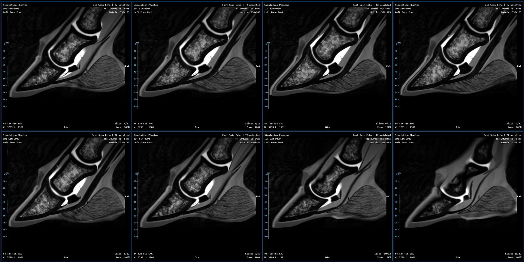
- Practice without the magnetSynthetic exam mode simulates a full acquisition on a modelled equine limb and reconstructs it through the same pipeline.
- Looks after itselfEvery system reports its operating state to Altra. Analysis of that data confirms the machine is behaving as it did at acceptance and flags drift early. Operating data only, never patient images.
How you buy it
No capital purchase
A scan and service fee puts a 0.4T system in your clinic for the cost of the cases it scans. Our interest and yours are the same: a system that is scanning.
Pay per scan
A fee per scan plus a service fee, instead of a capital outlay. Lower cost to install, and a cost that follows your caseload.
Always current
The service fee keeps the system on the current software release and under Altra’s care, with new sequences and features as they ship.
Watched over
Every system reports its own operating state to Altra. We usually know about a problem before you do. Operating data only, never patient images.
Sequence library
Every family, every plane, any angle
Sagittal, coronal and transverse as standard, and any oblique angle on the pilot. Field of view 170 mm; 12 slices per stack at 5 mm, or 3.5 mm in high-resolution variants.
| Family | Weightings | Variants |
|---|---|---|
| Spin echo | T1, proton density | Standard, high resolution |
| Fast spin echo | T2, proton density | Standard, fast, high resolution, super-fast, long TE |
| STIR fast spin echo | Fluid-sensitive, fat-suppressed | Four quality levels; selectable inversion time |
| Gradient echo 2D | T1, T2* (in and out of phase) | Standard, fast, high resolution |
| Gradient echo 3D | T1, T2* | Standard, high resolution, 1.0 mm isotropic |
| Dual echo | Proton density + T2 | Three planes |
| Pilot | Three-plane locator | Under one minute |
Specifications
What changes at 0.4T
Standing systems in service today
- Homogeneity
- Under 110 ppm over a 150 mm sphere
- Motion correction
- Navigator echo in fast spin echo and STIR, three samples per second, corrected in processing
- Console
- Dedicated Windows PC
- Remote access
- Third-party screen-sharing tool
From the manufacturer's published specification and product pages, 2025–2026.
AltraMRI
- Homogeneity
- 35 ppm over a 150 mm sphere
- Motion correction
- Continuous tracking, every sequence family
- Console
- Web application on iPad, laptop or any browser
- Remote access
- Native, through the browser
| Magnet | 0.4 T permanent magnet |
|---|---|
| Homogeneity / imaging volume | 35 ppm over a 150 mm sphere |
| Gradients | 20 mT/m |
| RF | 6 kW amplifier; four-channel digital spectrometer; integral transmit coil for a uniform B1 field |
| Coils | Hoof, fetlock and carpus limb receive coils |
| Anatomy | Fore and hind foot, pastern, fetlock and suspensory; carpus; tarsus |
| Field of view | 170 mm standard |
| Slices | 2D: 5 mm (3.5 mm HR), 12 per stack. 3D: 2.4 / 1.6 / 1.0 mm isotropic |
| Matrix | 170 × 170 acquired as standard (1.0 mm in plane at 170 mm field of view); up to 192 × 192 high resolution (0.9 mm in plane) |
| Patient position | Standing, sedated, forward-facing or rear-facing on the platform |
| Motion correction | Continuous limb tracking, independent of the pulse sequence, all sequence families |
| Console | AltraMRI web application: iPad, laptop or any modern browser on the clinic network |
| Reconstruction | GPU server; deep-learning denoise and de-Gibbs on complex data |
| Output | DICOM; PACS push (C-STORE); verified native archive with offline storage and restore |
| Access control | Operator, Clinical, Administrator and Demonstration roles |
| Training | Synthetic exam simulation, no magnet required |
| Commercial model | Scan and service fee. No capital purchase |
| Service | Connected health monitoring with operating data only; early systems delivered by trailer for on-site acceptance |
Background: gradient-coil design notes from the Altra engineering archive.
Request
The brochure, the manual, or a seat at the console
Tell us who you are and what you would like. Documents go out by email, usually within one working day. For a test drive we set you up with your own login to a console running in simulation, so you can plan and run studies on a synthetic horse from wherever you are.
Sales brochure
The system, the protocol, the commercial model. PDF.
Operator manual
How a study is run on the console, from positioning to export. PDF.
Console test drive
Remote access to the AltraMRI console in simulation, as a demonstration user. We send a private network invitation and a login; no magnet required.